Coronary Thrombosis vs Embolism: The Real Difference Behind Two Different Heart Attacks
Coronary Thrombosis vs. Embolism: What’s Really Blocking Your Heart Artery (And Why It Changes Everything)
Table of Contents
Picture two people rushed into the same cardiac cath lab on the same night. Both are having a heart attack. Both have the same crushing chest pain, the same sweating, the same alarming readings on the monitor. But when the cardiologist threads a catheter into their arteries and looks at the blockage on screen, the two cases look nothing alike.
One patient has a jagged, messy blockage sitting on top of years of built up plaque. The other has one clean, sharp stoppage in an artery that otherwise looks brand new. Same emergency room, same symptoms, same treatment team. Two completely different diseases causing it.
This is the difference between coronary thrombosis and coronary embolism. Most people, including a lot of patients who’ve actually had a heart attack, have never heard that these are two separate things. But understanding the difference changes what treatment looks like, what tests get ordered afterward, and what a patient needs to worry about down the road.
The One Sentence Difference You Need to Remember
Coronary thrombosis is a clot that forms right where it causes trouble. Coronary embolism is a clot that forms somewhere else in the body and travels to the heart.
That’s it. That’s the whole distinction, and everything else in this article is really just an expansion of that one idea.
In thrombosis, the artery itself is sick. It’s been building up fatty plaque for years, maybe decades, and the clot is the final, dramatic event on top of an already damaged blood vessel.
In embolism, the artery was probably fine seconds before the event happened. The clot started its life somewhere completely different, most often inside a heart chamber, on a heart valve, or even in a leg vein, and it simply got swept along by the bloodstream until it hit a spot narrow enough to get stuck.
Coronary Thrombosis vs Embolism: How Common Is Each One, Really?
If you had to bet on which one caused a random heart attack, bet on thrombosis every single time. It’s the cause behind the overwhelming majority of heart attacks treated worldwide.
Coronary embolism is the rare one. Research puts it behind roughly 3% to 7% of all acute coronary syndrome cases, and in some studies focused specifically on AF related cases, the number for atrial fibrillation driven coronary embolism has been reported as low as 2.9% of acute MI patients.
Here’s why that rarity actually matters clinically. Doctors are trained to expect thrombosis. It’s what they see day in and day out. So when a patient shows up with a clean, oddly located blockage and no real risk factors, it’s easy for that pattern to get missed or explained away as an unusual case of standard disease, rather than flagged as a completely different mechanism that needs a different workup.
Wait, Do They Feel Different?
No, and this is worth saying clearly because a lot of people assume there must be some telltale symptom that gives it away. There isn’t.
Chest pain, pressure, pain radiating down the arm or up into the jaw, shortness of breath, nausea, cold sweats. All of it is identical whether the blockage is homegrown thrombosis or an embolism that traveled in from somewhere else. The initial ECG changes and the troponin blood test results look the same too, at least at first.
The only way to tell them apart is by actually looking inside the artery during angiography, and sometimes even that isn’t enough on its own. Doctors often have to piece the picture together using something called the Shibata Criteria, imaging tests to hunt for where a clot might have originated, and occasionally a cardiac MRI to catch details that a standard angiogram misses.
What’s Actually Happening Inside a Thrombosis
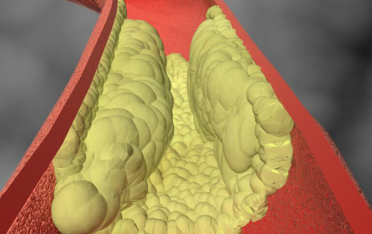
To understand thrombosis, you have to understand plaque. Fatty deposits build up inside artery walls over years, forming a bump called a plaque. Some plaques are stable and just sit there quietly. Others develop a thin, fragile covering that can tear open without warning.
There are two different ways this goes wrong, and the split between them is more interesting than most explanations give it credit for.
Plaque rupture is the classic version. The thin covering over a fatty plaque tears, and the gooey, fat rich material underneath gets exposed directly to flowing blood. Blood treats this exposed material as an emergency and immediately starts clotting on top of it, sometimes closing off the entire artery within minutes. Autopsy research has consistently found that plaque rupture accounts for around 55% to 60% of fatal coronary thrombosis cases, making it the single biggest driver of sudden coronary death.
Plaque erosion is the quieter, less talked about sibling. Here, the protective covering over the plaque never actually tears. Instead, the thin layer of cells lining the inside of the artery wears away, exposing a rougher surface that blood clots stick to anyway. A landmark study by Arbustini and colleagues found that plaque erosion was behind 25% of fatal heart attack cases overall, and it showed up far more often in women (37.4%) than in men (18.5%). Some more recent estimates push that number even higher, suggesting erosion might explain up to 40% of acute coronary syndrome cases, especially in younger patients, women, and smokers.
Why does this split matter beyond trivia? Because erosion related blockages sometimes behave differently once treated. Patients with plaque erosion tend to have better blood flow right after the event and respond well to aggressive blood thinning medication, which has led some researchers to ask whether certain erosion cases could be managed with medication alone, skipping a stent entirely. That question is still being actively studied, but it’s already reshaping how some cardiologists approach younger female patients with unusual sounding heart attacks.
Either way you slice it, rupture or erosion, the common thread stays the same. The artery itself was already unwell before the clot formed. The disease was building for years before the emergency ever happened.
What’s Actually Happening Inside an Embolism
Embolism plays by a completely different set of rules, because the clot doesn’t originate in the heart’s own blood vessels at all.
Atrial Fibrillation Is the Number One Culprit
If there’s one condition to know about when it comes to coronary embolism, it’s atrial fibrillation, commonly shortened to AF. When the heart’s upper chambers quiver chaotically instead of beating in a coordinated way, blood doesn’t get squeezed out efficiently. It pools instead, and it pools in one very specific pocket called the left atrial appendage, a small pouch shaped like a wrinkled finger hanging off the main chamber.
That pouch is dangerous specifically because blood moves so slowly through it. Detailed fluid dynamics research has confirmed that this pocket has dramatically lower blood flow and higher clot forming risk than the rest of the heart chamber, which is exactly why it’s the epicenter of clot formation during AF. In fact, more than 90% of clots that form in the atria during AF originate specifically in this pouch, rather than anywhere else in the chamber.
Once a clot forms there, it doesn’t take much for a piece to break free, travel down through the aorta, and get funneled straight into a coronary artery.
Other Sources Worth Knowing About
Atrial fibrillation gets most of the attention, and for good reason, but it isn’t the only source of coronary embolism. A few others come up regularly in the medical literature:
- Heart valve problems. Infected valves (a condition called endocarditis) can shed infected clot fragments. Mechanical replacement valves can also form clots if blood thinning isn’t managed carefully.
- Clots left behind after a previous heart attack. A weakened, poorly contracting section of heart muscle from an earlier event can develop its own clot, which can later break loose.
- Heart tumors. Rare, but a growth called a myxoma can shed small fragments that travel downstream.
- Patent foramen ovale. This one deserves its own explanation because it’s genuinely surprising the first time you hear about it.
The PFO Story: When a Leg Clot Ends Up in Your Heart
Nearly everyone is born with a small flap between the heart’s two upper chambers that’s supposed to close shortly after birth. In roughly a quarter of people, it never fully seals. Most of the time this causes zero problems and people live their entire lives never knowing it’s there.
But under the right circumstances, a clot that forms in a deep leg vein can travel up toward the lungs, and instead of getting filtered out the normal way, it can slip through that open flap and shoot straight into the arterial circulation. This is called a paradoxical embolism, and while most of the time it ends up causing a stroke, every so often it lands in a coronary artery instead.
One documented case involved a completely healthy 26 year old man who showed up with sudden chest pain and turned out to have a paradoxical embolism through a previously unknown PFO.
Doctors used aspiration thrombectomy to clear the blockage without placing a stent, since imaging confirmed there was no actual artery disease underneath. He later had the hole in his heart closed to prevent it from happening again.
Cases like this are genuinely rare. In broader research looking at paradoxical embolism overall, heart attack makes up a small minority of the outcomes, with most paradoxical emboli instead traveling to the brain and causing stroke. But when it does happen to land in a coronary artery, it’s a textbook example of why the source matters just as much as the blockage itself.
How Doctors Actually Tell the Two Apart
For a long time there was no formal system for making this call, just clinical judgment and hindsight from autopsy findings. That changed in 2015 when a group of Japanese researchers led by Dr. Shibata proposed a scoring system that’s now widely referred to as the NCVC criteria, named after Japan’s National Cerebral and Cardiovascular Center.
The system uses a mix of major and minor clues. The big ones are angiographic findings that look like embolism rather than typical plaque rupture, evidence of clots blocking more than one artery territory at once, and signs of clot fragments showing up elsewhere in the body at the same time. Supporting clues include a suspiciously clean looking artery everywhere except the blockage itself, an identifiable source visible on imaging like echocardiogram, and known risk factors such as atrial fibrillation.
Even with a formal scoring system in hand, this is genuinely hard to nail down in real time. In one study applying these criteria, doctors were only able to confirm embolism definitively during the procedure itself in 55.6% of suspected cases, while the other 44.4% couldn’t be fully verified through angiography or intervention alone. That’s a useful reality check. Even experienced interventional cardiologists sometimes have to make their best educated guess in the moment.
What the Blockage Actually Looks Like on Screen
This is often the first tip off a cardiologist gets, well before any formal scoring happens.
A thrombosis typically looks messy and irregular, sitting right on top of plaque that’s usually visible not just at that one spot but scattered throughout the rest of the artery too. It’s a local disaster happening inside a chronically diseased pipe.
An embolism typically looks clean. A smooth, round blockage or an abrupt, sharp cutoff, usually in an artery that looks otherwise pristine everywhere else. Case reports describing embolism again and again mention this same pattern, blockages showing up in branch vessels or further down the artery than typical plaque related blockages usually occur, sitting in a vessel with no other visible disease. This makes sense once you think about it mechanically. A traveling clot doesn’t stop where plaque happens to be. It stops wherever the pipe finally gets narrow enough to trap it, which is often further downstream than where homegrown blockages usually form.
A Blind Spot Worth Knowing About: MINOCA
Here’s a scenario that trips up a lot of people, patients and sometimes even clinicians early in training. Someone has all the classic signs of a heart attack: elevated troponin, abnormal ECG changes, damaged heart muscle on imaging. But when they get an angiogram, the arteries look basically fine. No major blockage anywhere.
This scenario has a name: MINOCA, which stands for Myocardial Infarction with Non-Obstructive Coronary Arteries. It’s more common than most people realize. A pooled analysis of over 800,000 patients found MINOCA showed up in about 8.1% of all heart attack cases, and individual national registries have reported numbers ranging anywhere from roughly 3% up to 15% depending on how strictly the diagnosis is defined.
It also skews heavily female. Women make up more than half of MINOCA cases, a striking reversal from standard obstructive heart disease, where men are the majority. MINOCA patients also tend to be younger and have fewer of the usual risk factors like diabetes, high cholesterol, or smoking.
Coronary embolism is one of the recognized causes hiding behind a MINOCA diagnosis, especially when a clot is small enough, or breaks apart and moves further downstream, before the angiogram gets a clear look at it. Other causes lurking behind a normal looking angiogram include coronary artery spasm, a condition called spontaneous coronary artery dissection where the artery wall itself tears without any plaque involved, and inherited clotting disorders. Screening for these clotting disorders in MINOCA patients has actually turned up meaningful numbers, with inherited conditions like factor V Leiden or protein C and S deficiency found in roughly 14% of patients tested.
For years MINOCA was assumed to be the “good” version of a heart attack, since the pipes looked clean. That assumption has aged poorly. Longer term studies now show MINOCA patients face real ongoing cardiovascular risk, with reported annual death rates as high as 5%, and outcomes that in some studies look statistically similar to patients with standard, obstructive coronary disease rather than meaningfully better. A clean angiogram after a heart attack should raise more questions, not fewer.
Why Treatment Actually Splits in Two Different Directions
This is where getting the diagnosis right stops being academic and starts changing what actually happens to the patient.
Treating Thrombosis
Since the artery itself is genuinely diseased, the standard playbook applies. A stent gets placed to physically prop the artery open and scaffold the unstable section of vessel wall. Patients go home on dual antiplatelet medication (usually aspirin plus a second blood thinning drug), a high dose statin to stabilize any remaining plaque, and a serious conversation about diabetes control, blood pressure, and smoking, since all of those directly feed the disease that caused the event in the first place.
Treating Embolism
Here the logic flips. If the artery itself has no disease, jamming a permanent metal stent into a perfectly healthy section of vessel doesn’t make sense. It solves nothing about why the clot showed up in the first place, and it leaves the patient with a foreign object sitting in an artery that never needed one, along with the long term medication burden that comes with it.
Instead, the preferred approach is aspiration thrombectomy, essentially vacuuming the clot straight out through the catheter and leaving the native artery untouched. This shows up again and again in real case reports. In one case involving a suspected paradoxical embolism, aspiration successfully restored blood flow and no stent was placed at all, since intravascular imaging confirmed there was no underlying artery disease to justify one. A similar case involving an 86 year old woman found the artery completely clear of disease after the clot was aspirated, so no stent was used.
It’s worth flagging that this actually runs a bit counter to general modern guidance. Current European Society of Cardiology guidelines have moved away from routinely recommending thrombectomy devices for standard heart attack care, mainly because in ordinary thrombosis, sucking out the clot doesn’t fix the ruptured plaque sitting underneath it. That guidance was built around thrombosis. In embolism specifically, the calculation is different, because there’s no plaque left behind to worry about once the clot is gone.
Beyond the procedure itself, treating an embolism properly means chasing down and treating whatever caused it in the first place:
- If AF caused it, the patient needs a blood thinner (an anticoagulant, not just aspirin or a standard antiplatelet drug), since these medications specifically target the kind of clot that forms in areas of pooled, slow moving blood.
- If a valve infection caused it, that means antibiotics, and sometimes surgery to repair or replace the valve.
- If a PFO caused it, the workup includes checking the legs for a hidden vein clot, screening for clotting disorders, and having a genuinely nuanced conversation about whether closing that hole in the heart makes sense. This isn’t always a clear cut decision. Current evidence on whether to close a PFO after a heart related paradoxical embolism is far less settled than the evidence supporting PFO closure after a stroke, so these calls tend to be made case by case rather than off a standard protocol.
Does One Carry a Worse Prognosis Than the Other?
There’s no clean, satisfying answer here like “embolism is always worse” or “thrombosis is always worse.” Outcomes really hinge on the specific source, how big the blockage was, and how quickly everything got identified and treated.
What the research does make clear is that a “clean” looking artery is not automatically the safer diagnosis. Comparisons between MINOCA patients and patients with standard obstructive disease have found broadly similar long term outcomes between the two groups, including comparable rates of repeat heart attacks, which pushes back hard against the old assumption that a normal angiogram automatically meant a low risk patient.
For coronary embolism specifically, the real long term danger usually isn’t the first event. It’s what happens if the underlying source never gets treated. An AF patient left on inadequate blood thinning, a valve infection that goes untreated, or a PFO left wide open in someone with an ongoing clotting disorder can all send another clot downstream, whether that lands in the heart again, the brain, or somewhere else entirely. This is exactly why chasing down and fixing the source matters just as much as clearing the immediate blockage.
Quick Reference: Thrombosis vs Embolism Side by Side
| What You’re Looking At | Coronary Thrombosis | Coronary Embolism |
|---|---|---|
| Where the clot is born | Right at the blockage site | Somewhere else (heart chamber, valve, leg vein) |
| Condition of the artery | Diseased, plaque filled | Usually normal, no disease |
| How common | The vast majority of heart attacks | Around 3% to 7% of ACS cases |
| Look on angiogram | Irregular, messy, plaque visible elsewhere too | Smooth, sharp, sudden cutoff, rest of artery looks clean |
| Where it typically strikes | Bigger, high pressure zones near branch points | Often further downstream, smaller branch vessels |
| Typical patient | Diabetes, high cholesterol, smoker, older | Often young, few risk factors, known AF or valve issue |
| Other clues | Usually isolated to the heart | Sometimes clot fragments show up elsewhere too (stroke, other organs) |
| First treatment choice | Stent placement | Aspiration to remove clot, often no stent |
| Long term medication | Antiplatelet drugs plus statin | Blood thinner (anticoagulant) plus treating the source |
| Symptoms | Identical | Identical |
The Bottom Line
If you strip away all the imaging jargon and diagnostic scoring systems, there’s a simplified version of this that holds up reasonably well in most real cases.
A messy, irregular blockage sitting on top of plaque scattered throughout the rest of the artery points toward thrombosis, and the standard heart attack playbook applies.
A single, clean, sharp cutoff in an otherwise healthy looking artery, in someone with atrial fibrillation, a heart valve problem, points toward embolism, and the next step becomes finding where that clot actually came from.
Neither pattern is bulletproof on its own. Small or distal clots can slip past even good imaging, and the diagnostic tools available today still have real limits.
But getting this distinction right is the difference between a patient walking away with a stent and a statin prescription nobody knew they had, a hidden valve infection, in the heart that had been quietly there since birth.